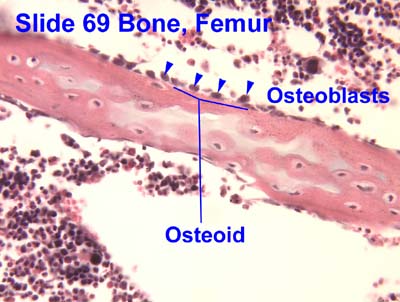
Osteoid
Template:Short description Template:More footnotes
Page Module:Infobox/styles.css has no content.
| Osteoid | |
|---|---|
| Lua error in package.lua at line 80: module 'Module:InfoboxImage/data' not found. | |
| Anatomical terminology |
Script error: No such module "Check for conflicting parameters".
In histology, osteoid is the unmineralized, organic portion of the bone matrix that forms prior to the maturation of bone tissue.[1] Osteoblasts begin the process of forming bone tissue by secreting the osteoid as several specific proteins. The osteoid and its adjacent bone cells have developed into new bone tissue when it becomes mineralized.[citation needed]
Osteoid makes up about fifty percent of bone volume and forty percent of bone weight. It is composed of fibers and ground substance. The predominant type of fiber is type I collagen and comprises ninety percent of the osteoid. The ground substance is mostly made up of chondroitin sulfate and osteocalcin.[citation needed] Osteoblasts synthesize and secrete osteoid as an unmineralized organic matrix, and when the osteoid becomes mineralized through deposition of calcium salts, it transforms into mature bone tissue.[2]
Composition
Osteoid is primarily composed of Type I collagen and ground substance.
Collagen fibers
Type I collagen comprises approximately 85-95% of the organic matrix,[3] providing the structural scaffold and tensile strength necessary for bone formation.[4] These collagen fibers form a dense, highly cross-linked network that serves as the foundation for subsequent mineralization.[5]
The collagen molecules are arranged in layers that alternate parallel and orthogonal to the axis of stress loading, creating a sophisticated composite structure.[5]
Ground substance
The ground substance of osteoid comprises approximately 10% of the bone matrix[6] and includes non-collagenous proteins:
- Proteoglycans - Including chondroitin sulfate and heparan sulfate, which bind to collagen and may regulate collagen fibril diameters and play a role in mineralization[3]
- Osteocalcin - A bone-specific protein involved in binding calcium during the mineralization process[3]
- Osteonectin - May serve a bridging function between collagen and the mineral component[3]
- Bone sialoprotein - Proteins rich in sialic acid that participate in matrix organization[2]
- Growth factors - Including transforming growth factors, fibroblast growth factors, and insulin-like growth factors[3]
Formation and secretion
Osteoid is synthesized and secreted by osteoblasts, specialized bone-forming cells.[7] Osteoblasts are large cuboidal cells characterized by abundant rough endoplasmic reticulum, reflecting their high synthetic activity.[4]
At their apical surface, osteoblasts secrete large amounts of type I collagen and smaller amounts of matrix organizing proteins, including osteocalcin and osteopontin.[5] The newly secreted osteoid forms a hydrated protein matrix layer between the mineralization front and the osteoblast layer.[5][7]
The synthesis of bone matrix occurs in two main steps: deposition of organic matrix (osteoid) and its subsequent mineralization.[7] During active bone formation, osteoblasts continuously produce osteoid matrix until mineralization occurs.
Mineralization
Script error: No such module "Labelled list hatnote".
The transformation of osteoid into mineralized bone involves the deposition of hydroxyapatite crystals [Page Module:Chem2/styles.css has no content.Ca10(PO4)6OH2] within the organic matrix.[6]
Mechanism of mineralization
Mineralization occurs in two phases: the vesicular phase and the fibrillar phase.[7]
Vesicular phase: Matrix vesicles, measuring 30-200 nm in diameter, are released from the apical membrane of osteoblasts into the newly formed bone matrix.[7][3] These vesicles contain alkaline phosphatase, adenosine triphosphatase (ATPase), and inorganic pyrophosphatase, and act as seeding sites for hydroxyapatite crystal formation through localized enzymatic accumulation of calcium and phosphate.[3]
Role of alkaline phosphatase: Osteoblasts secrete alkaline phosphatase, which participates in bone mineralization by hydrolyzing pyrophosphate, an inhibitor of mineralization, thereby increasing local inorganic phosphate availability for hydroxyapatite formation.[2][4]
Crystal propagation: Crystal growth proceeds from initial foci in matrix vesicles to form spheroids, which gradually coalesce to form a network of apatite crystals.[3] As the matrix matures, hydroxyapatite microcrystals are organized into a sophisticated composite within the collagen layer by nucleation in the protein lattice.[5]
Clinical significance
Disorders of osteoid mineralization
Defective mineralization of osteoid leads to several important clinical conditions characterized by accumulation of unmineralized matrix.
Rickets: Occurs in children when defective mineralization affects the growth plate and osteoid.[8] The condition results from calcium or phosphate deficiency, either isolated or secondary to vitamin D deficiency.[9] In rickets, the mineralization defect leads to accumulation of osteoid in bone tissue below the growth plate, resulting in weak bones and deformities.[9]
Osteomalacia: The adult equivalent of rickets, osteomalacia is characterized by softened bones due to impaired mineralization of osteoid.[10] Vitamin D deficiency is the most common cause, though calcium or phosphate deficiency can also result in osteomalacia.[10] The condition manifests as bone pain, muscle weakness, and increased fracture risk.
Histomorphometric characteristics of osteomalacia include:[11][9]
- Prolonged mineralization lag time (greater than 100 days)
- Widened osteoid seams (increased osteoid thickness)
- Increased osteoid volume
Vitamin D metabolism disorders: Rickets and osteomalacia develop in various clinical situations and have in common an absence or delay in the mineralization of growth cartilage and newly formed bone collagen.[12] Deficiency of vitamin D, essential for absorption of dietary calcium, has been a major cause historically.[12]
Osteoid in bone tumors
Osteosarcoma: The most common primary malignant bone tumor, osteosarcoma is characterized by malignant cells that produce osteoid.[13][14] Osteosarcoma is a primary malignant tumour of the skeleton characterized by the direct formation of immature bone or osteoid tissue by the tumour cells.[14]
The production of osteoid by tumor cells is the histological hallmark distinguishing osteosarcoma from other bone tumors.[15] The malignant osteoid produced appears as irregular, immature structures in close proximity to malignant cells with enlarged hyperchromatic nuclei and abnormal mitotic figures.[16]
Osteoid osteoma: A benign bone-forming tumor characterized by formation of osteoid tissue, typically causing localized bone pain that worsens at night.[17]
Role in bone remodeling
Osteoid plays a central role in the continuous process of bone remodeling.[2] During the bone formation phase of remodeling, osteoblasts deposit new osteoid at sites previously resorbed by osteoclasts.[7]
The bone remodeling cycle involves distinct phases:[2]
- Activation: Recruitment of osteoclasts to specific bone sites
- Resorption: Osteoclastic removal of old bone
- Reversal: Transition from resorption to formation
- Formation: Osteoblastic deposition of new osteoid
- Mineralization: Transformation of osteoid into mineralized bone
Throughout this process, osteoblasts produce the unmineralized organic matrix that subsequently undergoes calcification to form new bone tissue.[4]
Biochemical markers
Several biochemical markers are associated with osteoid metabolism and bone formation:
- Alkaline phosphatase: Secreted by osteoblasts during active bone formation; elevated levels indicate increased osteoid production[2][9]
- Osteocalcin: A bone-specific protein synthesized by osteoblasts and incorporated into osteoid; serves as a marker of bone formation[5]
- Type I collagen peptides: Degradation products measured to assess bone turnover
Physiology
Page Module:Message box/ambox.css has no content.
![[icon]](/wiki/File:Wiki_letter_w_cropped.svg) | This section needs expansion. You can help by Template:Protected page maintenance message. (June 2025) |
To allow bone mineralization to take place, osteoblasts secrete tissue-nonspecific alkaline phosphatase into the osteoid to break down pyrophosphate, an extracellular inhibitor of hydroxyapatite precipitation which otherwise prevents precipitation of hydroxyapetite crystals from extracellular fluid which is supersaturated in Ca2+ and Page Module:Chem2/styles.css has no content.PO3−4 ions.[18]
Disorders
When there are insufficient nutrient minerals or osteoblast dysfunction, the osteoid does not mineralize properly and accumulates. The resultant disorder is termed rickets in children and osteomalacia in adults. A deficiency of type I collagen, such as in osteogenesis imperfecta, also leads to defective osteoid and brittle, fracture-prone bones.
In some cases, secondary hyperparathyroidism can cause a disturbance in mineralisation of calcium and phosphate.
Another condition is a disturbance in primitive transformed cells of mesenchymal origin, which exhibit osteoblastic differentiation and produce malignant osteoid. This results in the formation of a malignant primary bone tumor known as osteosarcoma or osteogenic sarcoma. This malignancy most often develops in adolescence during periods of rapid osteoid formation (commonly referred to as growth spurts).[19]
See also
- Bone
- Bone matrix
- Osteoblast
- Osteocyte
- Bone mineralization
- Hydroxyapatite
- Rickets
- Osteomalacia
- Osteosarcoma
- Alkaline phosphatase
References
Page Template:Reflist/styles.css has no content.
- ^ Page Module:Citation/CS1/styles.css has no content.Trammell, Lindsay H.; Kroman, Anne M. (2013-01-01), DiGangi, Elizabeth A.; Moore, Megan K. (eds.), "Chapter 13 - Bone and Dental Histology", Research Methods in Human Skeletal Biology, Academic Press, pp. 361–395, doi:10.1016/b978-0-12-385189-5.00013-3, ISBN 978-0-12-385189-5, retrieved 2020-11-18
{{citation}}: CS1 maint: work parameter with ISBN (link) - ^ a b c d e f Page Module:Citation/CS1/styles.css has no content.White, H. J.; Helwany, M.; Biknevicius, A. R.; Peterson, D. C. (2024). "Physiology, Bone". StatPearls. StatPearls Publishing. PMID 30855919.
- ^ a b c d e f g h Page Module:Citation/CS1/styles.css has no content.Othman, D. (2012). "An Overview of Bone Cells and their Regulating Factors of Differentiation". Plastic Surgery International. 2012. doi:10.1155/2012/374398. PMC 3341892. PMID 22701169.
- ^ a b c d Cite error: The named reference
histology_osteoblastswas invoked but never defined (see the help page). - ^ a b c d e f Page Module:Citation/CS1/styles.css has no content.Blair, H. C.; Larrouture, Q. C.; Li, Y.; Lin, H.; Beer-Stoltz, D.; Liu, L.; Tuan, R. S.; Robinson, L. J.; Schlesinger, P. H.; Nelson, D. J. (2017). "Osteoblast Differentiation and Bone Matrix Formation In Vivo and In Vitro". Tissue Engineering Part B: Reviews. 23 (3): 268–280. doi:10.1089/ten.TEB.2016.0454. PMC 5467150. PMID 27846781.
- ^ a b Page Module:Citation/CS1/styles.css has no content.Han, D.; Wang, T.; Wang, R.; Chen, J.; Tang, Y. (2023). "A Brief Review of Bone Cell Function and Importance". Diagnostics. 13 (22). doi:10.3390/diagnostics13223443. PMC 10648520. PMID 37998580.
- ^ a b c d e f Page Module:Citation/CS1/styles.css has no content.Sun, S.; Huang, S. Q. (2015). "Biology of Bone Tissue: Structure, Function, and Factors That Influence Bone Cells". International Journal of Clinical and Experimental Medicine. 8 (5): 8254–8258. PMC 4515490. PMID 26221406.
- ^ Page Module:Citation/CS1/styles.css has no content.Baron, S. L.; Mathai, J. K. (2024). "Rickets". StatPearls. StatPearls Publishing. PMID 32809510.
- ^ a b c d Page Module:Citation/CS1/styles.css has no content.Ghorpade, D. S.; Leyland, R.; Kurowska-Stolarska, M.; Patil, S. A.; Balaji, K. N. (2012). "Rickets–vitamin D deficiency and dependency". Molecular and Cellular Biology. 32 (12): 2239–2253. doi:10.1128/MCB.06597-11. PMC 3313732. PMID 22473996.
- ^ a b Page Module:Citation/CS1/styles.css has no content.Majmundar, V. D.; Syed, H. A.; Baxi, K. (2024). "Osteomalacia". StatPearls. StatPearls Publishing. PMID 32644463.
- ^ Page Module:Citation/CS1/styles.css has no content.Uday, S.; Högler, W. (2021). "Nutritional rickets & osteomalacia: A practical approach to management". The Indian Journal of Medical Research. 152 (4): 356–367. doi:10.4103/ijmr.IJMR_1961_19. PMC 8061584. PMID 33380700.
- ^ a b Page Module:Citation/CS1/styles.css has no content.Berry JL, Davies M, Mee AP (2002). "Vitamin D metabolism, rickets, and osteomalacia". Seminars in Musculoskeletal Radiology. 6 (3): 173–82. doi:10.1055/s-2002-36714. PMID 12541194.
{{cite journal}}: CS1 maint: multiple names: authors list (link) - ^ Page Module:Citation/CS1/styles.css has no content.Rishor-Olney, C. R.; Hinson, M. R. (2024). "Osteosarcoma (Osteogenic Sarcoma)". StatPearls. StatPearls Publishing. PMID 32644347.
- ^ a b Page Module:Citation/CS1/styles.css has no content.Fink-Retter, A.; Gschwantler-Kaulich, D.; Hudelist, G.; Mueller, R.; Kubista, E.; Czerwenka, K.; Singer, C. F. (2006). "Osteosarcoma (Osteogenic sarcoma)". Oncology Reports. 18 (2): 299–304. PMC 1794406. PMID 17611648.
- ^ Page Module:Citation/CS1/styles.css has no content.Dahlin DC, Coventry MB (1967). "Pathology of osteosarcoma". Clinical Orthopaedics and Related Research. 111 (111): 23–32. doi:10.1097/00003086-197509000-00004. PMID 168999.
- ^ Page Module:Citation/CS1/styles.css has no content."Osteosarcoma". Holland-Frei Cancer Medicine - NCBI Bookshelf. BC Decker. 2003.
- ^ Page Module:Citation/CS1/styles.css has no content.Exo, J.; Smith, C.; Smith, R.; Bell, M. (2010). "Update on Bone Forming Tumors of the Head and Neck". Pediatric Health. 3 (6): 533–541. doi:10.2217/phe.09.54. PMC 2807498. PMID 20191093.
- ^ Page Module:Citation/CS1/styles.css has no content.Hall, John E.; Hall, Michael E. (2021). "Chapter 55: Spinal Cord Motor Functions; the Cord Reflexes". Guyton and Hall Textbook of Medical Physiology (14th ed.). Philadelphia, PA: Elsevier. pp. 994–995. ISBN 978-0-323-59712-8.
- ^ Page Module:Citation/CS1/styles.css has no content.Ottaviani Giulia; Jaffe Norman (2009). "The Epidemiology of Osteosarcoma". Pediatric and Adolescent Osteosarcoma. Cancer Treatment and Research. Vol. 152. New York: Springer. pp. 3–13. doi:10.1007/978-1-4419-0284-9_1. ISBN 978-1-4419-0283-2. PMID 20213383.
- Page Module:Citation/CS1/styles.css has no content.Netter, Frank H. (1987), Musculoskeletal system: anatomy, physiology, and metabolic disorders, Summit, New Jersey: Ciba-Geigy Corporation
- Page Module:Citation/CS1/styles.css has no content.Jaffe, N.; et al. (2009). Pediatric and Adolescent Osteosarcoma. New York: Springer. ISBN 978-1-4419-0283-2.
External links
- Histology image: 69_03 at the University of Oklahoma Health Sciences Center - "Bone, femur"
- Dr. Susan Ott's website on osteomalacia
Lua error in package.lua at line 80: module 'Module:Navbox/configuration' not found.